Advances in Microscopy: A Gateway to Enhanced Research, Drug Development, and Diagnostics
Unlocking New Frontiers in Microscopy: Enhancing Research, Drug Development, and Diagnostics
Bruker’s Acquifer IM (Imaging Machine) is a fully automated widefield microscope ideal for high-content screening assays and phenotypic screening for small-model organisms.
Its static sample holder and moving optics renders it ideal for imaging motion-sensitive samples, such as non-adherent cell cultures or embryos.
In this webinar, Prof. Nicole Endlich will give an overview of her research on kidney disease and the development of new diagnostic tools. She will speak on:
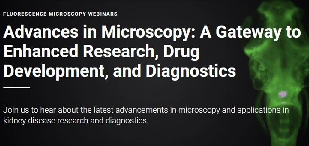
Developing high-content screening methods based on a zebrafish larvae model for the identification of potential therapeutic agents
Combing super resolution-based imaging techniques with multiplex staining and in situ hybridization for the advanced visualization of structure and morphology
Prof. Nicole Endlich’s talk offers an insightful exploration into the latest advancements in the microscopy technology and their transformative applications in scientific research, drug development, and diagnostics. A focal point of the presentation is the adoption of zebrafish larvae as an efficient model organism in high-content screening, specifically tailored for kidney research. This model facilitates the effective screening of potential pharmaceuticals. In addition, the presentation will cover innovative, super resolution-based imaging techniques that surpass the capabilities of traditional light microscopy. These techniques, combined with multiplex staining and in situ hybridization, allow the visualization of structures previously undetectable, marking the beginning of a new era in both research methodologies and diagnostic procedures.
Presenter: Prof. Nicole Endlich (Greifswald University Medicine, Germany)
Prof. Nicole Endlich’s talk offers an insightful exploration into the latest advancements in the microscopy technology and their transformative applications in scientific research, drug development, and diagnostics. A focal point of the presentation is the adoption of zebrafish larvae as an efficient model organism in high-content screening, specifically tailored for kidney research. This model facilitates the effective screening of potential pharmaceuticals. In addition, the presentation will cover innovative, super resolution-based imaging techniques that surpass the capabilities of traditional light microscopy. These techniques, combined with multiplex staining and in situ hybridization, allow the visualization of structures previously undetectable, marking the beginning of a new era in both research methodologies and diagnostic procedures.