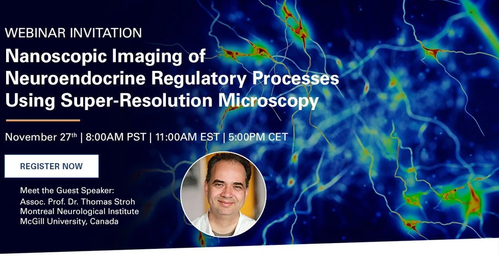
Nanoscopic Imaging of Neuroendocrine Regulatory Processes Using Super-Resolution Microscopy

Join us and our special guest speaker Dr. Thomas Stroh, Assistant Professor at Montreal Neurological Institute, McGill University, Canada, for this webinar on Nanoscopic Imaging of Neuroendocrine Regulatory Processes Using Super-Resolution Microscopy. Dr. Thomas Stroh is a leading researcher in the field of G protein-coupled receptors. His work focuses on studying the mechanisms governing the availability of receptors at the cell surface of neurons and of neuroendocrine cells, factors that can ultimately determine their sensitivity to transmitters, neuropeptides, and active agents.
Don’t miss the opportunity to hear Dr. Stroh give an overview of his research and innovative work on:
- Using the super-resolution microscopy techniques dSTORM (direct Stochastic Optical Reconstruction Microscopy) and biplane SMLM (single molecule localization microscopy) to image neuroendocrine regulatory processes in the brain at the near-molecular level
- Combining super-resolution microscopy with plasma hormone level testing to study receptor trafficking and synaptic remodeling in neuroendocrine circuits in pituitary cells and brain tissue
- Quantification of synaptic remodeling under the influence of hormonal secretion cycles in mouse brains
Presenter: Thomas Stroh, Ph.D. (Associate Professor, Montreal Neurological Institute, McGill University)
Assoc. Prof. Thomas Stroh has over 20 years of experience working in the fields of microscopy and neurobiology. In 2005, he established the Neuro Microscopy Core Facility of the Montreal Neurological Institute (The Neuro) of McGill University, which has since grown into a modern microscopy platform housing advanced microscopy ranging from live cell microscopy to fluorescence super-resolution microscopy. It currently serves >50 laboratories from The Neuro, various McGill departments, other Montreal universities and industry. Dr. Stroh has taught in numerous workshops and microscopy summer schools, including the Montreal Light Microscopy Course and the Canadian Light Microscopy Course at the University of Calgary. Together with Dr. Claire Brown of McGill University’s Advanced BioImaging Facility (ABIF), he organized the first Canadian Quantitative Super-Resolution Microscopy Course at The Neuro in 2019. Dr. Stroh is on the Executive Board of the Canada BioImaging Network. His research focuses on studying the regulation and trafficking of G Protein-Coupled Receptors. More recently, the Stroh lab is combining highly sensitive plasma hormone level testing with super-resolution microscopy to study the effects of endogenous rhythms on synaptic connectivity and plasticity in the hypothalamus.
Presenter's Abstract
Nanoscopic Imaging of Neuroendocrine Regulatory Processes in the Hypothalamo-Pituitary Axis Using dSTORM
Single molecule localization microscopy (SMLM) using direct Stochastic Optical Reconstruction Microscopy (dSTORM) enables the imaging of regulatory processes in the brain at the near-molecular level, which cannot be resolved using traditional fluorescence microscopy techniques. However, imaging brain samples is challenging due to the optical density of the samples. Nevertheless, suitable tissue clearing, biplane imaging to capture 3D data, as well as computational and statistical methods to differentiate between signal and noise can overcome these difficulties.
Here, we used biplane SMLM and dSTORM in combination with highly sensitive determination of plasma hormone levels to investigate receptor trafficking and synaptic remodeling within the hypothalamo-pituitary endocrine circuit in endocrine pituitary cells and brain tissue, respectively. In corticotropic pituitary cells, we identified a novel syntaxin-6 positive compartment that is distinct from the trans-Golgi-network (TGN) by dSTORM. This compartment facilitates the regulated recycling of the somatostatin receptor subtype 2 (SSTR2) to the cell surface following Corticotropin-Releasing Hormone stimulation, using nearest-neighbor analysis to determine the size of this compartment. In the hypothalamus, we used dSTORM to image synaptic connectivity and its remodeling in sections through the arcuate nucleus of the mouse hypothalamus in parallel with the pulsatile rhythm of Growth Hormone (GH) secretion, employing biplane imaging and the DBSCAN clustering algorithm. We observed that during peak GH secretion, there were more excitatory synapses at the somata of GH-releasing hormone (GHRH) neurons.
Conversely, during periods of low GH levels, inhibitory synaptic input increased on GHRH cells. The results presented here highlight biplane imaging and dSTORM as a valuable quantitative approach to study receptor trafficking and synaptic structure in neuroendocrine circuits. Using appropriate sample preparation, SMLM imaging techniques, and careful statistical analysis we achieved a nanometer-scale precision in identifying a novel compartment in the pituitary cells as well as imaging and quantifying synaptic remodeling under the influence of hormonal secretion cycles in mouse brain.
